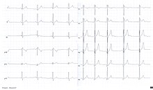

The U wave is a wave on an electrocardiogram (ECG). It comes after the T wave of ventricular repolarization and may not always be observed as a result of its small size. 'U' waves are thought to represent repolarization of the Purkinje fibers.[1][2] However, the exact source of the U wave remains unclear. The most common theories for the origin are:
- Delayed repolarization of Purkinje fibers
- Prolonged re-polarisation of mid-myocardial M-cells
- After-potentials resulting from mechanical forces in the ventricular wall
- The repolarization of the papillary muscle.[3]
- ^ Pérez Riera AR, Ferreira C, Filho CF, et al. (2008). "The enigmatic sixth wave of the electrocardiogram: the U wave". Cardiol J. 15 (5): 408–21. PMID 18810715.
- ^ "ECG Learning Center - An introduction to clinical electrocardiography". ecg.utah.edu. Retrieved 2017-01-02.
- ^ F., Boron, Walter; L., Boulpaep, Emile (2012). Medical physiology : a cellular and molecular approach. ISBN 9781437717532. OCLC 756281854.
{{cite book}}: CS1 maint: multiple names: authors list (link)
